
The ophthalmologist evaluates the patient, agrees with your initial exam. He confirms a diagnosis of acute angle closure glaucoma.
The ophthalmologist also attempts gonioscopy, but the view is poor through the edematous cornea.
What’s gonioscopy?
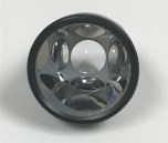 A Gonioscopy lens (image on left) has mirrors which allows for the examiner to visualize the angle of the eye, or the area where the trabecular meshwork is located, and where aqueous outflow occurs. In an eye with an open angle, the trabecular meshwork should be visible, as seen below:
 Above Image Credit License: CC-BY SA 3.0
However, gonioscopic exam of a patient with angle closure, with a clearer view, through less edematous cornea, would look like this:

In this patient, the iris is pushed up against the angle occluding the view of the trabecular meshwork, and therefore obstructing aqueous from the leaving the eye.
The ophthalmologist measures the patient’s glasses and notes that the patient is hyperopic with the following prescription:
OD +5.00 sph
OS: +4.50 sph
He orders aqueous suppressant drops and walks away to find a consent form.
Click here to move on!
Case 8 Index
Case 8: Introduction
Case 8: Physical Exam